Trajectory SP906
Force field:
martini_v2.2
Simulation length (ns): 5000
Electric field (kJ mol-1 nm-1 e-1): 0
Temperature (K): 300 (v-rescale)
Pressure (bar): 1 (Parrinello-rahman semiisotropic)
Number of particles: 19192
Time step (fs) : 25
Software: GROMACS 2021.5
Simulation length (ns): 5000
Electric field (kJ mol-1 nm-1 e-1): 0
Temperature (K): 300 (v-rescale)
Pressure (bar): 1 (Parrinello-rahman semiisotropic)
Number of particles: 19192
Time step (fs) : 25
Software: GROMACS 2021.5
Supercomputer:
Finisterrae III CESGA
Peptides: P243 NC03728
Lipids: POPC
Heteromolecules:
Ions: CL
Water model: W
Peptides: P243 NC03728
Lipids: POPC
Heteromolecules:
Ions: CL
Water model: W
Sequence :
RGGRLSYSRRRFSTSTGR
Total charge (e): +6
Number of residues: 18
By amino acid: Basic: 6 Acidic: 0 Hydrophobic: 5 Polar: 7 Electrostatic Dipolar Moment (e nm): 5.33
Longitudinal (e nm): 5.17 Transversal (e nm): 1.32 Hydrophobic Dipolar Moment (nm): 0.66
Longitudinal (nm): 0.22 Transversal (nm): 0.62 Secondary structure: Helix
Activity:
Download Files
ITP file. JSON file. PDB file.
Click on any component to highlight it from the plot.
Upper leaflet
Lower leaflet
Lipids
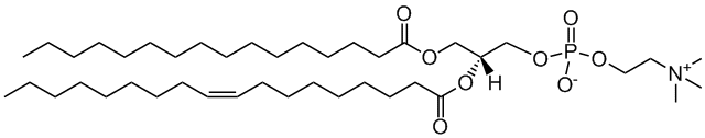
Membrane model for: POPC (Healthy mammal)
POPC
2-oleoyl-sn-glycero-3-phosphocholine
Total charge (e): 0
See POPC lipid
Download ITP File. Download PDB File.
Last snapshot
Total contacts per residue
Molecular Dynamics based descriptors
Average and standard deviation,
calculated using the autocorrelation function (for time
series)
or the width of the distribution, for the last microsecond
of
the trajectory
Area per lipid
Membrane (nm2): 0.642518000 ± 0.000985121
Upper leaflet (nm2): 0.642518000 ± 0.000985121
Lower leaflet (nm2): 0.642518000 ± 0.000985121
Average Z coordinate
Peptide (nm): 4.6869200 ± 0.0311067
First Residue (nm): 4.7973300 ± 0.0376575
Last Residue (nm): 4.5506900 ± 0.0536494
Membrane (nm): 6.8349400 ± 0.0102061
Upper leaflet Head Group (nm): 8.7962700 ± 0.0122452
Lower leaflet Head Group (nm): 4.87369000 ± 0.00836607
Bilayer Thickness (nm): 3.9225800 ± 0.0148302
Peptide insertion (nm): 0.186773 ± 0.032212
Contacts
Peptide - Water: 35.745000 ± 0.796451
Peptide - Head groups: 13.755000 ± 0.279967
Peptide - Tail groups: 7.282500 ± 0.271145
Tilt (°): 89.82000 ± 1.45641
PepDF:
5(ns): CVS
Displacement (nm): 0.667820 ± 0.026835
Precession(°): -1.24898 ± 1.83969
50(ns) CVS
Displacement (nm): 2.0345700 ± 0.0915866
Precession(°): -9.49259 ± 5.22491
100(ns) CVS
Displacement(nm): 3.018010 ± 0.141916
Precession(°): -19.61000 ± 5.74149
200(ns) CVS
Displacement(nm): 4.284610 ± 0.216002
Precession(°): -47.54010 ± 7.54271
Download JSON File.
Peptide Analyses
Peptide Displacement Fingerprint
(PepDF)
Lateral displacement vs
Rotational
Displacement along the trajectory, for different time
windows .
Density maps:
2D-density maps of lipids around the
peptide
along XY and YZ axis, calculated for each lipid type along the
last
microsecond.
Lipid-Peptide Analyses:
z-Position
Z-coordinate, averaged for
differetn
parts of the the system: peptide, membrane, first and
last
backbone (BB) residues and upper of lower leaflet
lipids’
headgroups (HGs).
Minimum distance
Minimum distance (nm) between the
peptide backbone and the lipids (headgroups and
tailgroups).
Number of contacts
Number of contacts between the
peptide backbone and the water or the lipids separated
by
lipid headgroups (HG) or lipid tails, using a cut-off of
0.6
nm.
Lateral density
Lateral density for the different
components of the system: headgroups, tail groups,
peptide
and water.