Trajectory SP754
Force field:
martini_v2.2
Simulation length (ns): 5000
Electric field (kJ mol-1 nm-1 e-1): 0
Temperature (K): 300 (v-rescale)
Pressure (bar): 1 (Parrinello-rahman semiisotropic)
Number of particles: 19183
Time step (fs) : 25
Software: GROMACS 2021.5
Simulation length (ns): 5000
Electric field (kJ mol-1 nm-1 e-1): 0
Temperature (K): 300 (v-rescale)
Pressure (bar): 1 (Parrinello-rahman semiisotropic)
Number of particles: 19183
Time step (fs) : 25
Software: GROMACS 2021.5
Supercomputer:
Finisterrae III CESGA
Peptides: P167 AP03166
Lipids: POPC
Heteromolecules:
Ions: CL
Water model: W
Peptides: P167 AP03166
Lipids: POPC
Heteromolecules:
Ions: CL
Water model: W
Sequence :
FSSLFKAGAKYLLKQVGKAGAQQLACKAANNC
Total charge (e): +5
Number of residues: 32
By amino acid: Basic: 5 Acidic: 0 Hydrophobic: 17 Polar: 10 Electrostatic Dipolar Moment (e nm): 6.75
Longitudinal (e nm): 6.5 Transversal (e nm): 1.82 Hydrophobic Dipolar Moment (nm): 6.94
Longitudinal (nm): 6.87 Transversal (nm): 1 Secondary structure: Helix
Activity:
Download Files
ITP file. JSON file. PDB file.
Click on any component to highlight it from the plot.
Upper leaflet
Lower leaflet
Lipids
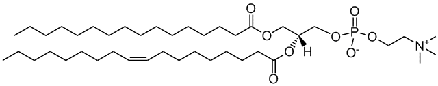
Membrane model for: POPC (Healthy mammal)
POPC
2-oleoyl-sn-glycero-3-phosphocholine
Total charge (e): 0
See POPC lipid
Download ITP File. Download PDB File.
Last snapshot
Total contacts per residue
Molecular Dynamics based descriptors
Average and standard deviation,
calculated using the autocorrelation function (for time
series)
or the width of the distribution, for the last microsecond
of
the trajectory
Area per lipid
Membrane (nm2): 0.644687000 ± 0.000956842
Upper leaflet (nm2): 0.644687000 ± 0.000956842
Lower leaflet (nm2): 0.644687000 ± 0.000956842
Average Z coordinate
Peptide (nm): 8.6389600 ± 0.0308823
First Residue (nm): 8.5807700 ± 0.0460002
Last Residue (nm): 8.7869100 ± 0.0442682
Membrane (nm): 6.80262000 ± 0.00974329
Upper leaflet Head Group (nm): 8.7622500 ± 0.0117551
Lower leaflet Head Group (nm): 4.8464900 ± 0.0078875
Bilayer Thickness (nm): 3.9157600 ± 0.0141561
Peptide insertion (nm): -0.1232900 ± 0.0330439
Contacts
Peptide - Water: 38.012500 ± 0.718822
Peptide - Head groups: 18.232500 ± 0.311404
Peptide - Tail groups: 16.74250 ± 0.33654
Tilt (°): 87.994300 ± 0.710896
PepDF:
5(ns): CVS
Displacement (nm): 0.6073740 ± 0.0266575
Precession(°): -0.0923751 ± 1.0378400
50(ns) CVS
Displacement (nm): 1.9160700 ± 0.0903641
Precession(°): -1.53836 ± 2.47642
100(ns) CVS
Displacement(nm): 2.911960 ± 0.126827
Precession(°): -0.149717 ± 2.925910
200(ns) CVS
Displacement(nm): 3.239840 ± 0.153884
Precession(°): 1.50895 ± 3.15446
Download JSON File.
Peptide Analyses
Peptide Displacement Fingerprint
(PepDF)
Lateral displacement vs
Rotational
Displacement along the trajectory, for different time
windows .
Density maps:
2D-density maps of lipids around the
peptide
along XY and YZ axis, calculated for each lipid type along the
last
microsecond.
Lipid-Peptide Analyses:
z-Position
Z-coordinate, averaged for
differetn
parts of the the system: peptide, membrane, first and
last
backbone (BB) residues and upper of lower leaflet
lipids’
headgroups (HGs).
Minimum distance
Minimum distance (nm) between the
peptide backbone and the lipids (headgroups and
tailgroups).
Number of contacts
Number of contacts between the
peptide backbone and the water or the lipids separated
by
lipid headgroups (HG) or lipid tails, using a cut-off of
0.6
nm.
Lateral density
Lateral density for the different
components of the system: headgroups, tail groups,
peptide
and water.