Trajectory SP1366
Force field:
martini_v3
Simulation length (ns): 5000
Electric field (kJ mol-1 nm-1 e-1): 0
Temperature (K): 300 (v-rescale)
Pressure (bar): 1 (Parrinello-rahman semiisotropic)
Number of particles: 19211
Time step (fs) : 25
Software: GROMACS 2021.5
Simulation length (ns): 5000
Electric field (kJ mol-1 nm-1 e-1): 0
Temperature (K): 300 (v-rescale)
Pressure (bar): 1 (Parrinello-rahman semiisotropic)
Number of particles: 19211
Time step (fs) : 25
Software: GROMACS 2021.5
Supercomputer:
Finisterrae III CESGA
Peptides: P472 NC00130
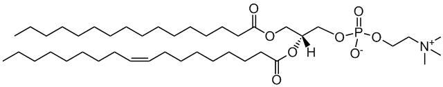
Lipids: POPC
Heteromolecules:
Ions: CL
Water model: W
Peptides: P472 NC00130
Lipids: POPC
Heteromolecules:
Ions: CL
Water model: W
Sequence :
LNVPPSWFLSQR
Total charge (e): +1
Number of residues: 12
By amino acid: Basic: 1 Acidic: 0 Hydrophobic: 7 Polar: 4 Electrostatic Dipolar Moment (e nm): 1.46
Longitudinal (e nm): 1.27 Transversal (e nm): 0.72 Hydrophobic Dipolar Moment (nm): 2.1
Longitudinal (nm): 2.07 Transversal (nm): 0.32 Secondary structure: Helix
Activity:
Download Files
ITP file. JSON file. PDB file.
Click on any component to highlight it from the plot.
Upper leaflet
Lower leaflet
Lipids
Membrane model for: POPC (Healthy mammal)
POPC
2-oleoyl-sn-glycero-3-phosphocholine
Total charge (e): 0
See POPC lipid
Download ITP File. Download PDB File.
Last snapshot
Total contacts per residue
Molecular Dynamics based descriptors
Average and standard deviation,
calculated using the autocorrelation function (for time
series)
or the width of the distribution, for the last microsecond
of
the trajectory
Area per lipid
Membrane (nm2): 0.64821700 ± 0.00104108
Upper leaflet (nm2): 0.64821700 ± 0.00104108
Lower leaflet (nm2): 0.64821700 ± 0.00104108
Average Z coordinate
Peptide (nm): 8.9369600 ± 0.0345985
First Residue (nm): 8.7210100 ± 0.0393463
Last Residue (nm): 9.389630 ± 0.047706
Membrane (nm): 6.8425200 ± 0.0107275
Upper leaflet Head Group (nm): 8.7860800 ± 0.0129667
Lower leaflet Head Group (nm): 4.89897000 ± 0.00861474
Bilayer Thickness (nm): 3.8871100 ± 0.0155676
Peptide insertion (nm): 0.1508820 ± 0.0369486
Contacts
Peptide - Water: 29.480000 ± 0.706071
Peptide - Head groups: 7.782500 ± 0.218895
Peptide - Tail groups: 6.570000 ± 0.295336
Tilt (°): 67.81110 ± 1.77125
PepDF:
5(ns): CVS
Displacement (nm): 0.9217110 ± 0.0374106
Precession(°): 4.72795 ± 4.84708
50(ns) CVS
Displacement (nm): 3.000100 ± 0.130819
Precession(°): 49.5894 ± 18.3965
100(ns) CVS
Displacement(nm): 4.302410 ± 0.183353
Precession(°): 97.8568 ± 24.8263
200(ns) CVS
Displacement(nm): 6.43429 ± 0.23632
Precession(°): 146.0810 ± 33.4724
Download JSON File.
Peptide Analyses
Peptide Displacement Fingerprint
(PepDF)
Lateral displacement vs
Rotational
Displacement along the trajectory, for different time
windows .
Density maps:
2D-density maps of lipids around the
peptide
along XY and YZ axis, calculated for each lipid type along the
last
microsecond.
Lipid-Peptide Analyses:
z-Position
Z-coordinate, averaged for
differetn
parts of the the system: peptide, membrane, first and
last
backbone (BB) residues and upper of lower leaflet
lipids’
headgroups (HGs).
Minimum distance
Minimum distance (nm) between the
peptide backbone and the lipids (headgroups and
tailgroups).
Number of contacts
Number of contacts between the
peptide backbone and the water or the lipids separated
by
lipid headgroups (HG) or lipid tails, using a cut-off of
0.6
nm.
Lateral density
Lateral density for the different
components of the system: headgroups, tail groups,
peptide
and water.